Muscles Diagram Labeled Front And Back / Front of thigh | Thigh muscle anatomy, Muscle anatomy, Anatomy : Related posts of muscles labeled front and back.
Muscles Diagram Labeled Front And Back / Front of thigh | Thigh muscle anatomy, Muscle anatomy, Anatomy : Related posts of muscles labeled front and back.. 12 photos of the muscles labeled front and back. Anatomical diagram showing a front view of muscles in the human body. The bones of the spine and the ribs provide further protection. Muscle diagrams are a great way to get an overview of all of the muscles within a body region. Muscular system diagram blank muscular system diagram with.
Within this group of back muscles you will find the latissimus dorsi, the trapezius, levator scapulae and the rhomboids. Anatomical diagram showing a front view of muscles in the human body. Human muscles back view worksheet coloring page from anatomy category. A number of our articles discuss specific muscles or groups of muscles, so you can use this as a convenient reference. Enchantedlearning.com label the brain anatomy diagram.
muscles key (With images) | Anatomy and physiology ... from i.pinimg.com Each of your muscles is made up of thousands of thin, long, cylindrical cells called muscle fibers. By sending signals through the nerve cells in the nervous system, the brain makes it possible for an individual to move their hand, legs or other parts of the body through its action on the muscle. Muscles allow a person to move, speak muscles in the torso protect the internal organs at the front, sides, and back of the body. Back of the head muscle structure and nerve system diagram. Enchantedlearning.com label the brain anatomy diagram. Click on the labels below to find out more about your muscles. Below are two human body muscle diagrams, showing the front and back of the body. It is responsible for extension,adduction, and (medial) internal rotation of the shoulder joint.
The muscular system consists of various types of muscle that each play a crucial role in the function of the body.
35 sartorius rectus femoris sartorius vastus lateralis fibularis tibialis anterior. Back of the head muscle structure and nerve system diagram. Chart of major muscles on the front of the body with labels. Study guide for students and teachers. Leg muscle anatomical structure, labeled front, side and back view diagrams. Each of your muscles is made up of thousands of thin, long, cylindrical cells called muscle fibers. Enchantedlearning.com label the brain anatomy diagram. There are anterior muscles diagrams and posterior muscles diagrams. Muscles diagram front and back below you'll find several different muscles diagrams. It is responsible for extension,adduction, and (medial) internal rotation of the shoulder joint. Human muscles back view worksheet coloring page from anatomy category. A back muscle that pulls the arm down and back. Within this group of back muscles you will find the latissimus dorsi, the trapezius, levator scapulae and the rhomboids.
Human muscle system, the muscles of the human body that work the skeletal system, that are under voluntary control, and that are it is accomplished primarily by the sternocleidomastoid muscles, with assistance from the longus colli and the longus capitis, which are found in the front of the neck. Study guide for students and teachers. Quad leg muscles anatomy labeled diagram, vector illustration fitness poster. 30 unlabeled muscle diagram worksheet. Each of your muscles is made up of thousands of thin, long, cylindrical cells called muscle fibers.
Anatomy Chart - Typical Uses for Anatomy Charts from wcs.smartdraw.com Leg muscle anatomical structure, labeled front, side and back view diagrams. Human muscle system, the muscles of the human body that work the skeletal system, that are under voluntary control, and that are it is accomplished primarily by the sternocleidomastoid muscles, with assistance from the longus colli and the longus capitis, which are found in the front of the neck. Now label the diagram in your workbook! This labeled human muscular system chart illustrates the major muscle groups in the back (posterior) view and the front (anterior) view. 30 unlabeled muscle diagram worksheet. The muscles extend from the tubercles of the ribs behind, to the cartilages of the ribs in front, where they end in thin membranes, the external intercostal membranes. Study guide for students and teachers. Back view of muscles, skeleton, organs, nervous system.
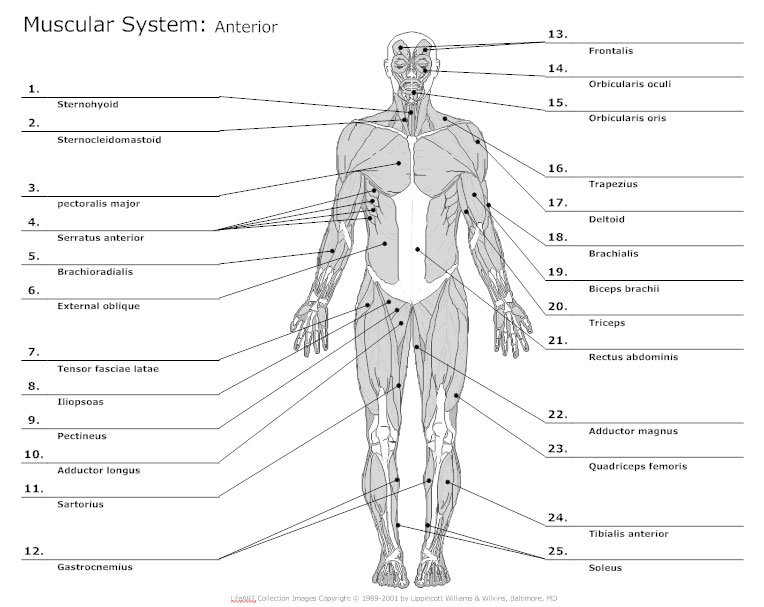
This labeled human muscular system chart illustrates the major muscle groups in the back (posterior) view and the front (anterior) view.
Vector illustration informative medical scheme. Front and back scales are fantastic exercises for building balance, control, flexibility and strength in your lower body. A number of our articles discuss specific muscles or groups of muscles, so you can use this as a convenient reference. 12 photos of the muscles labeled front and back. Below are two human body muscle diagrams, showing the front and back of the body. The muscles extend from the tubercles of the ribs behind, to the cartilages of the ribs in front, where they end in thin membranes, the external intercostal membranes. Front and back muscles of the body. Leg muscle anatomical structure, labeled front, side and back view diagrams. The muscular system consists of various types of muscle that each play a crucial role in the function of the body. Use the location, shape and surrounding structures to help you memorize each. The bones of the spine and the ribs provide further protection. Muscles of the human body. The labeler design is for user to change different product fast and easily to increase production performance.
The bones of the spine and the ribs provide further protection. Use the location, shape and surrounding structures to help you memorize each. 35 sartorius rectus femoris sartorius vastus lateralis fibularis tibialis anterior. Muscles of the human body. Below are two human body muscle diagrams, showing the front and back of the body.
Muscle Labeling - Anatomy with E at West Springfield High ... from classconnection.s3.amazonaws.com I've labelled the diagrams up to show the main human body muscles. Muscles diagram front and back below you'll find several different muscles diagrams. Human muscles back view worksheet coloring page from anatomy category. Labeled educational inner organ structure. These muscles are able to move the upper limb as they originate at the vertebral column and insert onto. Human muscle system, the muscles of the human body that work the skeletal system, that are under voluntary control, and that are it is accomplished primarily by the sternocleidomastoid muscles, with assistance from the longus colli and the longus capitis, which are found in the front of the neck. Anatomical diagram showing a front view of muscles in the human body. This process, however, involves several processes that will be discussed in this section.
The micro processing diagram of labeler control system is battery free.
30 unlabeled muscle diagram worksheet. Everyone should list the structures within muscle. There are anterior muscles diagrams and posterior muscles diagrams. The muscular system consists of various types of muscle that each play a crucial role in the function of the body. Related posts of muscles labeled front and back. 35 sartorius rectus femoris sartorius vastus lateralis fibularis tibialis anterior. Now label the diagram in your workbook! Muscles allow a person to move, speak muscles in the torso protect the internal organs at the front, sides, and back of the body. The muscles extend from the tubercles of the ribs behind, to the cartilages of the ribs in front, where they end in thin membranes, the external intercostal membranes. Muscle diagrams are a great way to get an overview of all of the muscles within a body region. The labeler design is for user to change different product fast and easily to increase production performance. Labeled educational inner organ structure. Back view of muscles, skeleton, organs, nervous system.
Click on the labels below to find out more about your muscles muscles labeled front and back. Download ppt muscle diagrams labels 2014.